


Mitophagy is the process your cells use to identify and remove damaged mitochondria before they cause problems. It’s one of the most important quality-control mechanisms in the body, and research shows it gradually slows down as you age.
Understanding mitophagy is useful for anyone serious about longevity. The state of your mitochondria has a direct influence on cellular energy, tissue resilience, brain function, and how well your body handles the demands of daily life.
Key Takeaways

The Short Version
Mitophagy is a cellular recycling process that clears out damaged mitochondria and allows the cell to replace them with healthier ones.
The primary pathway that triggers mitophagy involves two proteins called PINK1 and Parkin, which detect membrane damage and signal for removal.
Mitophagy declines measurably with age, and that decline is associated with lower energy output, reduced muscle function, and poor cellular maintenance across multiple tissues.
Exercise, calorie restriction, and certain compounds including Urolithin A have been shown in human studies to activate mitophagy pathways.
What Is Mitophagy?
Mitophagy is the selective degradation of mitochondria through autophagy. Mitochondria generate the energy your cells run on, but they accumulate damage over time from oxidative stress, replication errors, and normal wear. Mitophagy is the mechanism cells use to identify faulty mitochondria, break them apart, and recycle the components.
The word itself comes from “mito” (mitochondria) and “phagy” (eating), a reference to the lysosomal digestion process that dismantles the targeted organelle.
A 2023 review published in Nature Metabolism describes mitophagy as a key regulator of mitochondrial quality, noting that it also supports the biogenesis of new, functional mitochondria by clearing the old ones out of the way. The system isn’t just disposal — it’s renewal.
How Mitophagy Works: The PINK1–Parkin Pathway
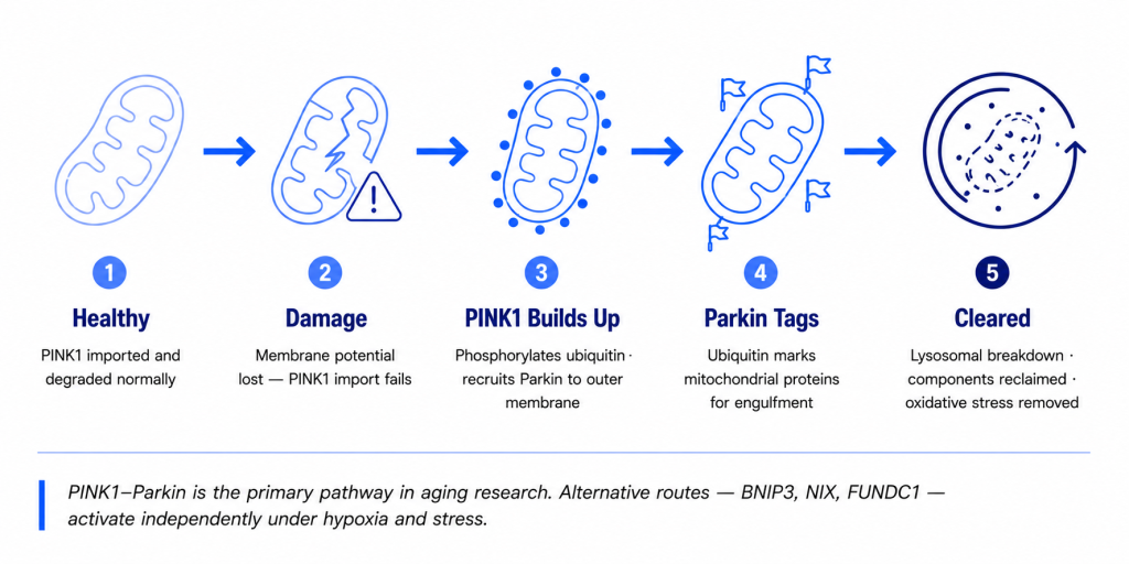
The best-characterized mitophagy pathway centers on two proteins: PINK1 (PTEN-induced kinase 1) and Parkin, a ubiquitin E3 ligase.
Under normal conditions, PINK1 is imported into healthy mitochondria and rapidly degraded. When a mitochondrion becomes damaged and loses its membrane potential, that import fails. PINK1 accumulates on the outer mitochondrial membrane, where it phosphorylates ubiquitin chains and recruits Parkin to the surface. Parkin then tags mitochondrial proteins with ubiquitin, flagging the organelle for autophagic engulfment and lysosomal breakdown.
The result: the damaged mitochondrion is consumed, its components are reclaimed for cellular use, and the cell has one fewer source of oxidative stress and metabolic inefficiency.
A review in Ageing Research Reviews outlines how PINK1/Parkin pathway disruption allows damaged mitochondria to accumulate in neurons, contributing to the cellular dysfunction seen in several age-related neurological conditions.
The PINK1–Parkin pathway is not the only route. Receptors including BNIP3, NIX, and FUNDC1 can initiate mitophagy independently of Parkin under hypoxia and other stress conditions. But PINK1–Parkin remains the primary pathway studied in aging and disease research.
Why Mitophagy Declines as You Age
Mitophagy is not a fixed constant. It’s a dynamic process that responds to cellular stress, energy status, and the efficiency of the lysosomal machinery that carries out the breakdown.
Evidence consistently shows that mitophagy slows with age. The same 2023 Nature Metabolism review documents measurably reduced mitophagy across multiple tissues in aging organisms, including decreases in the hippocampus, heart tissue, and skeletal muscle. In human skeletal muscle, mitochondrial function declines significantly with age, and there’s evidence that this has downstream effects on strength and physical capacity.
Several factors drive the decline. Lysosomal activity decreases with age, meaning the breakdown machinery becomes less efficient even when a mitochondrion is correctly tagged. NAD+ levels fall over time, and research published in Cold Spring Harbor Perspectives in Medicine notes that NAD+ supports mitophagy signaling through sirtuin-dependent pathways, meaning its decline compounds the problem. PINK1 and Parkin expression also appear to decrease in aged tissues.
The practical consequence: damaged mitochondria accumulate in cells at a rate the declining mitophagy machinery can no longer match.
What Happens When Damaged Mitochondria Accumulate
A buildup of dysfunctional mitochondria isn’t just an energy problem. Damaged mitochondria produce excess reactive oxygen species, amplifying oxidative stress inside the cell. They also leak mitochondrial DNA into the cytoplasm, which can trigger inflammatory signaling cascades.
A review published in Cells describes how age-related declines in autophagy and mitophagy allow toxic aggregates and damaged organelles to build up in neurons, contributing to progressive cellular dysfunction. The energy-intensive nature of neuronal tissue makes the brain particularly sensitive to mitochondrial quality control failures.
Beyond the brain, impaired mitophagy has been linked to changes in muscle metabolism, immune cell function, and cardiac tissue health. These effects show up at the tissue and organ level as the kinds of changes that are typically attributed to “normal aging” — declining energy, reduced physical resilience, slower recovery.
This is why researchers have characterized mitophagy not just as a cellular maintenance process, but as a meaningful target for healthy aging strategies.
How to Support Mitophagy: Lifestyle Inputs
Several well-studied lifestyle interventions activate mitophagy pathways without pharmaceutical intervention.
Exercise is one of the strongest triggers. Endurance exercise in particular upregulates mitophagy-related proteins in skeletal muscle, and this appears to be a key mechanism through which regular training supports mitochondrial quality over time.
Calorie restriction and fasting reliably induce mitophagy across multiple tissues. A literature review published in the Journal of Cachexia, Sarcopenia and Muscle examined the evidence across study types and found that caloric restriction consistently upregulates mitophagy markers including BNIP3 and Parkin. Time-restricted eating may offer a practical implementation path for people who want the benefit without sustained caloric deficit.
Sleep matters here too. Mitophagy appears to increase during sleep in certain brain regions, which may partly explain why chronic poor sleep accelerates neurological aging.
These lifestyle inputs are foundational. Supplement strategies work best when they’re layered on top of them, not instead of them.
Urolithin A and Mitophagy: What the Human Evidence Shows
Urolithin A is the best-studied nutritional compound for mitophagy activation in humans. It’s a postbiotic metabolite produced in the gut from ellagitannins, polyphenols found in pomegranates, walnuts, and certain berries. Because gut microbiome composition varies considerably between individuals, most people don’t produce meaningful amounts of Urolithin A from food alone, which is what has driven interest in supplementation.
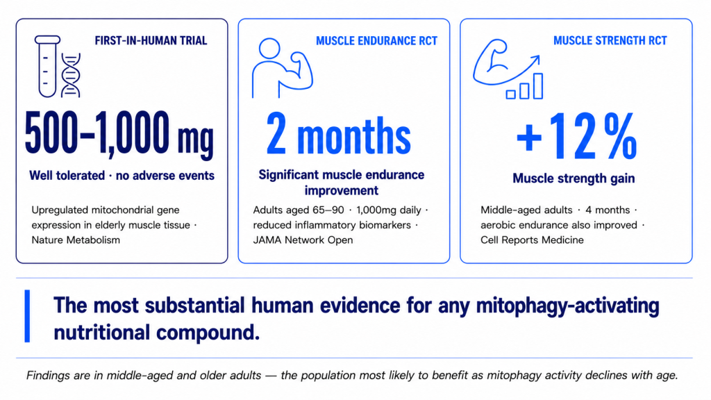
The first-in-human clinical trial, published in Nature Metabolism, found that four weeks of daily Urolithin A supplementation at 500 mg and 1,000 mg modulated plasma acylcarnitines and upregulated mitochondrial gene expression in skeletal muscle of elderly participants. The compound was well tolerated at all doses tested, with no significant adverse events.
A randomized controlled trial in JAMA Network Open enrolled adults aged 65 to 90 and found that daily supplementation with 1,000 mg of Urolithin A significantly improved muscle endurance at two months compared to placebo, alongside reductions in several inflammatory biomarkers and acylcarnitines associated with mitochondrial dysfunction.
A second randomized controlled trial published in Cell Reports Medicine examined middle-aged adults and found roughly 12% improvements in muscle strength alongside meaningful aerobic endurance improvements with Urolithin A supplementation over four months. The researchers also observed increases in muscle proteins linked to mitophagy and mitochondrial metabolism, suggesting the compound’s effects were mechanistically connected to the mitophagy pathway rather than being a nonspecific energy boost.
This is currently the most substantial body of human clinical evidence for a mitophagy-activating nutritional compound. The findings are in middle-aged and older adults, which is the population most likely to benefit from mitophagy support given the age-related decline in baseline activity.
What to Look for in a Mitophagy-Support Supplement
If you’re evaluating supplements for mitophagy support, a few factors actually matter.
Ingredient form. Urolithin A needs to be present as the active compound, not as a precursor. Ellagitannins from food don’t reliably convert to Urolithin A because that conversion depends on specific gut bacteria most people don’t have in sufficient quantities.
Dose. The human trials showing meaningful effects used 500 mg to 1,000 mg of Urolithin A daily. Formulas that include it as a minor ingredient in a larger blend are unlikely to reach these amounts.
Testing documentation. As with any longevity supplement, third-party testing and Certificates of Analysis confirm that the labeled compound is present at the labeled dose. The human trial data on Urolithin A is strong enough that cutting corners on verification undercuts the whole argument for taking it.
BioLongevity’s BioRecharge formula includes 1,000 mg of Urolithin A per serving alongside C60 fullerene, pomegranate extract, and alpha-lipoic acid, designed to support mitophagy and broader mitochondrial antioxidant defense as part of a daily longevity routine.
Frequently Asked Questions
What is the difference between mitophagy and autophagy?
Autophagy is a broad cellular recycling process that targets a range of damaged or unnecessary components, including proteins, lipids, and organelles. Mitophagy is a specific subtype of autophagy focused exclusively on mitochondria. Think of autophagy as the general housekeeping system and mitophagy as the specialized branch that handles the cell’s power plants.
Can you have too much mitophagy?
Yes, in theory. Excessive mitophagy, where too many functional mitochondria are removed, can impair energy production and is being studied in the context of certain pathological states. In practice, lifestyle interventions and nutritional compounds that activate mitophagy appear to work within a physiologically appropriate range. The concern with aging is almost always the opposite direction: insufficient mitophagy.
How long does it take for mitophagy support to show measurable effects?
The Urolithin A RCTs saw significant improvements in muscle endurance biomarkers at two months and muscle strength changes at four months of daily supplementation. Mitophagy-related changes at the gene and protein expression level were visible earlier. There’s no shortcut timeline, but the data suggests consistent daily use over multiple months is the right frame.
Does Urolithin A only affect muscle?
No. Muscle is where most of the clinical trial data exists because it’s a practical and measurable tissue. But Urolithin A activates PINK1–Parkin-dependent mitophagy pathways that operate across cell types. Preclinical research has examined mitophagy activation in neural tissue, and this is an active area of clinical investigation.
Is mitophagy the same as cellular senescence?
No. Cellular senescence is a state in which a cell stops dividing but doesn’t die. Mitophagy is a quality-control process that happens within cells, including senescent ones. Mitochondrial dysfunction is often present in senescent cells and can amplify the inflammatory signals those cells emit, which is one reason mitophagy is studied alongside senescence in aging research.
Can you directly measure mitophagy?
Not routinely in clinical settings. Researchers use specialized reporters and mitochondrial protein turnover assays in research contexts. In human trials, mitophagy activation is typically inferred from indirect markers: plasma acylcarnitines, muscle mitochondrial gene expression, and mitophagy-related protein levels. These are the same biomarkers that improved in the Urolithin A trials.
Mitophagy is one of the more mechanistically supported targets in longevity science. The pathway is well characterized, the age-related decline is documented, and the human evidence for nutritional support is more robust than for most longevity-adjacent ingredients. If you’re building a daily routine around cellular health, this is a mechanism worth understanding.
Ask a qualified clinician before starting any supplement if you are pregnant, nursing, taking medication, or managing a medical condition.
References
[1] Picca A, Faitg J, Auwerx J, Ferrucci L, D’Amico D. Mitophagy in human health, ageing and disease. Nat Metab. 2023;5(12):2047–2061. doi:10.1038/s42255-023-00930-8
[2] Singh A, D’Amico D, Andreux PA, et al. Urolithin A improves muscle strength, exercise performance, and biomarkers of mitochondrial health in a randomized trial in middle-aged adults. Cell Rep Med. 2022;3(5):100633. doi:10.1016/j.xcrm.2022.100633
[3] Liu S, D’Amico D, Shankland E, et al. Effect of Urolithin A Supplementation on Muscle Endurance and Mitochondrial Health in Older Adults: A Randomized Clinical Trial. JAMA Netw Open. 2022;5(1):e2144279. doi:10.1001/jamanetworkopen.2021.44279
[4] Andreux PA, Blanco-Bose W, Ryu D, et al. The mitophagy activator urolithin A is safe and induces a molecular signature of improved mitochondrial and cellular health in humans. Nat Metab. 2019;1(6):595–603. doi:10.1038/s42255-019-0073-4
[5] Li J, Yang D, Li Z, et al. PINK1/Parkin-mediated mitophagy in neurodegenerative diseases. Ageing Res Rev. 2022;84:101817. doi:10.1016/j.arr.2022.101817
[6] Mehrabani S, Bagherniya M, Askari G, Read MI, Sahebkar A. The effect of fasting or calorie restriction on mitophagy induction: a literature review. J Cachexia Sarcopenia Muscle. 2020;11(6):1447–1458. doi:10.1002/jcsm.12611
[7] Lautrup S, Hou Y, Fang EF, Bohr VA. Roles of NAD+ in Health and Aging. Cold Spring Harb Perspect Med. 2024;14(1). doi:10.1101/cshperspect.a041193
[8] Metaxakis A, Ploumi C, Tavernarakis N. Autophagy in Age-Associated Neurodegeneration. Cells. 2018;7(5). doi:10.3390/cells7050037